Case #154 – April, 2005
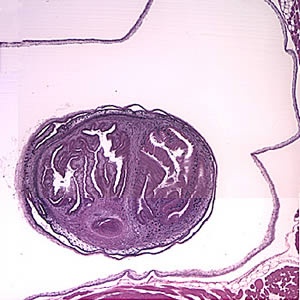
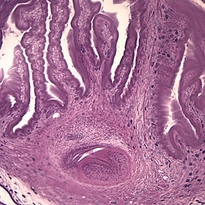
A 49-year-old male immigrant from Mexico was seen at a local medical facility specializing in neural disorders for frequent headaches and occasional seizures. Figures A and B show what was observed on a hematoxylin and eosin (H & E) stained section of lesions detected in the right frontal lobe of his brain. Figure A was taken at 40× magnification and Figure B was taken at 100× magnification. What is your diagnosis? Based on what criteria?

Figure A

Figure B
Images presented in the DPDx case studies are from specimens submitted for diagnosis or archiving. On rare occasions, clinical histories given may be partly fictitious.

