Case #161 – August, 2005
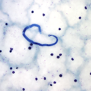
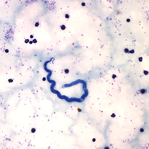
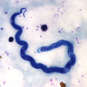
A 50-year-old returned to the United States after working with a missionary group in Mali and Senegal for 10 years. He went to a local hospital with a low-grade fever for 2 days. The physician ordered a blood film examination and thick and thin films were made and stained with Giemsa. Figures A-D show the objects, ranging in size from 236 to 245 micrometers, that were seen on the thick blood film. Figures A and C were captured at 400× magnification and Figures B and D were captured at 1000× magnification. What is your diagnosis? Based on what criteria?

Figure A

Figure B

Figure C

Figure D
Images presented in the DPDx case studies are from specimens submitted for diagnosis or archiving. On rare occasions, clinical histories given may be partly fictitious.