Case #182 – June, 2006
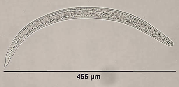
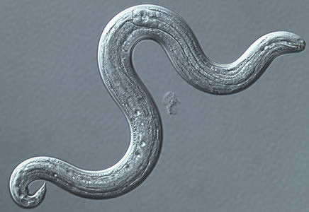
A researcher was studying parasites of public health concern found in snails and slugs. A small portion of one specimen had tissue removed and placed in a small dish with a HCl/pepsin solution. Many larvae were observed in the dish, however most were dead or dying, but a few larvae were active. Figure A shows one of the active larvae in at wet mount captured at 200× magnification. Figure B shows a larva under differential interference contrast (DIC) microscopy at 400× magnification. What is your identification of the objects? Based on what criteria?

Figure A

Figure B
Images presented in the DPDx case studies are from specimens submitted for diagnosis or archiving. On rare occasions, clinical histories given may be partly fictitious.

