Case #188 – September, 2006
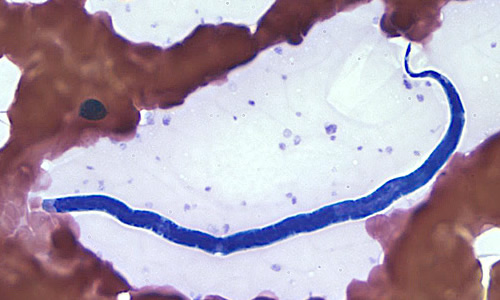
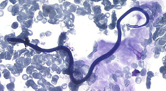
A hospital submitted blood films to CDC’s reference laboratory for identification of the microfilariae that were seen on the films. The object in Figure A was seen on a Wright-Giemsa stained blood film and measured approximately 180 µm. The object in B was seen on a Giemsa stained blood film and measured approximately 240-250 µm. Both A and B were taken at 500× magnification. What is your diagnosis? Based on what criteria?

Figure A

Figure B
Images presented in the DPDx case studies are from specimens submitted for diagnosis or archiving. On rare occasions, clinical histories given may be partly fictitious.
