Case #195 – January, 2007
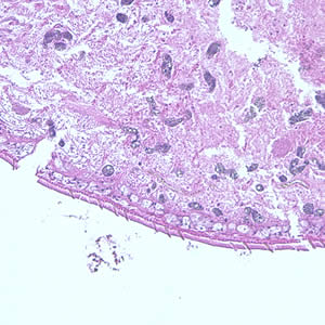
A patient complaining of intermittent symptoms including coughing up blood, fever, and other vague “flu-like” symptoms saw a physician. The patient reported working at a sushi restaurant and eating a raw crab on a dare (Figure A shows a crab similar to the one that the patient ate). Blood tests were ordered and results included peripheral eosinophilia of 10% and a history of bilateral pneumothorax (free air or gas in the pleural cavity). A biopsy yielded a cyst containing a structure 5 mm in length and 2 mm in width. Figure B (40×) and Figure C (100×) show a hematoxylin and eosin (H & E) stained section of the specimen. Figure D (400×) shows an object which measured 80-90 µm by 40-45 µm. Similar objects were found in low numbers in sections of lung tissue. What is your diagnosis? Based on what criteria?

Figure A

Figure B

Figure C

Figure D
Images presented in the DPDx case studies are from specimens submitted for diagnosis or archiving. On rare occasions, clinical histories given may be partly fictitious.


