Case #236 – September, 2008
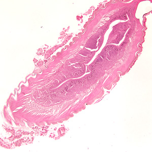
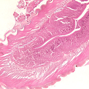
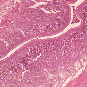
A 30-year-old who frequents sushi restaurants started experiencing severe gastritis, including epigastric pain, nausea and vomiting. He had reported eating at a sushi restaurant the previous day. After being admitted to the hospital for severe pain, a gastric biopsy was performed. A tissue specimen was sectioned and stained with hematoxylin and eosin (H&E). The attending pathologist observed unusual structures from the biopsied material and sent the slide to the CDC for diagnostic assistance. Figures A–C show structures observed on the slide; images were captured at 100x, 200x and 400x, respectively. What is your diagnosis? Based on what criteria?

Figure A

Figure B

Figure C
Images presented in the DPDx case studies are from specimens submitted for diagnosis or archiving. On rare occasions, clinical histories given may be partly fictitious.

