Case #275 – May, 2010
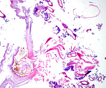
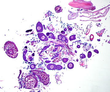
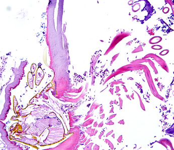
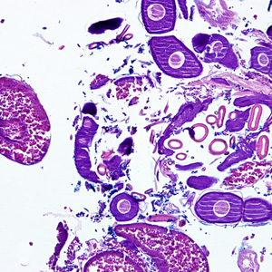
A 70-year-old female, who had recently returned from a trip to Madagascar, went to the hospital for a painful sensation on the underside of her left foot while walking. Examination of the area between the hallux and index toes revealed an ulcerative lesion. A biopsy was performed and sent to the Pathology Department for work-up. The specimen was sectioned, stained with hematoxylin and eosin (H&E) and examined by the attending pathologist. Figures A and B show what was observed at 40x magnification. Figures C and D show the same fields at 200x magnification, respectively. What is your diagnosis? Based on what criteria?

Figure A

Figure B

Figure C

Figure D
Images presented in the DPDx case studies are from specimens submitted for diagnosis or archiving. On rare occasions, clinical histories given may be partly fictitious.

