Case #366 – February 2014
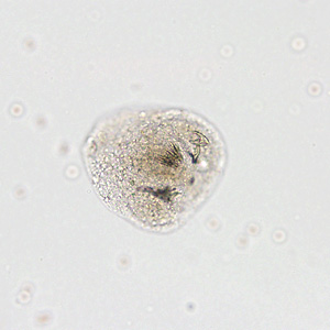
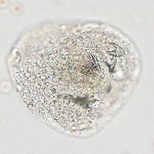
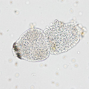
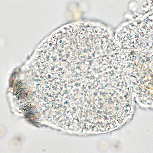
A 28-year-old male from Algeria had complaints of right upper quadrant (RUQ) abdominal pain. As part of his work up at a medical facility, imaging studies revealed a complex solid and cystic lesion in the posterior right hepatic dome. Hepatic cyst fluid was aspirated and submitted for ova-and-parasite (O&P) testing. Figures A–D show what was observed on a wet-mount made from the sediment of the fluid. Figures A and C were captured at 100x magnifications; Figures B and D at 200x magnification. What is your diagnosis? Based on what criteria?

Figure A

Figure B

Figure C

Figure D
Images presented in the DPDx case studies are from specimens submitted for diagnosis or archiving. On rare occasions, clinical histories given may be partly fictitious.