Case #374 – June 2014
A 34-year-old missionary worker sought medical attention for abdominal pain, nausea, and watery diarrhea after returning from visiting friends in Central America. A stool specimen was collected for laboratory testing and a modified Kinyoun’s acid-fast stained smear was prepared. An aliquot of the stool, along with the acid-fast stained smear, was sent to the CDC-DPDx laboratory for confirmatory testing. Figures A and B show what was observed in moderate numbers on the acid-fast stained smear at 500x magnification with oil. Figures C-F were taken at 400x magnification from a wet mount of the stool specimen. Figures C and E were taken using bright-field microscopy; Figures D and F represent the same fields, respectively, viewed using ultraviolet (UV) microscopy. The objects of interest measured 8-9 micrometers in diameter on average. What is your diagnosis? Based on what criteria?
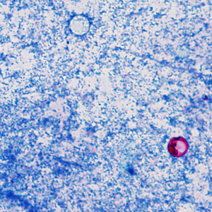
Figure A
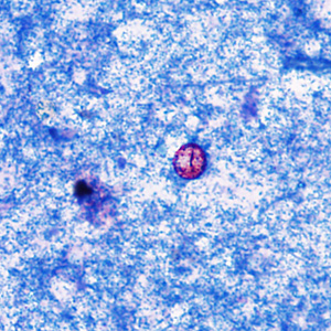
Figure B
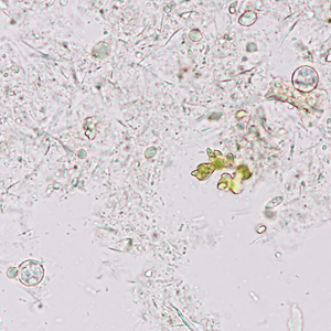
Figure C
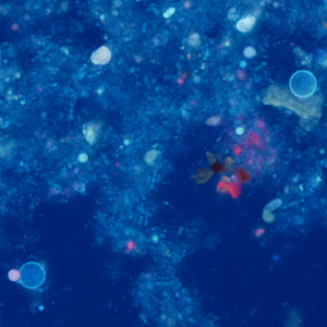
Figure D
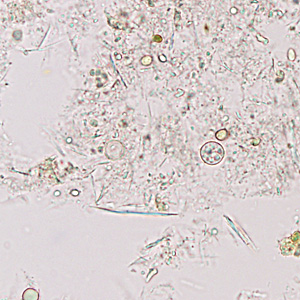
Figure E
Images presented in the DPDx case studies are from specimens submitted for diagnosis or archiving. On rare occasions, clinical histories given may be partly fictitious.