Case #401 – August 2015
A worm approximately three centimeters long was observed and removed during a routine colonoscopy of a 54-year-old man from Scandinavia. The worm was sent to the CDC-DPDx Team for identification. Figure A shows the gross worm at 10x magnification using a dissecting microscope. It was then cleared with lacto-phenol and examined at 100x magnification (Figures B and C show the anterior and posterior respectively). A thin cross-section was made and examined at the same magnification (Figure D) and 200x (Figure E). What is your diagnosis? Based on what criteria?
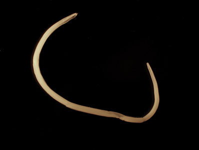
Figure A
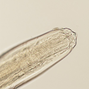
Figure B
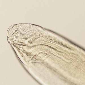
Figure C
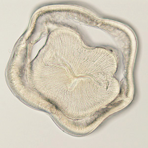
Figure D
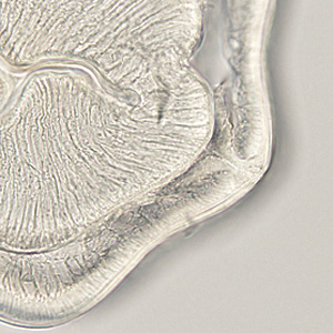
Figure E
Images presented in the DPDx case studies are from specimens submitted for diagnosis or archiving. On rare occasions, clinical histories given may be partly fictitious.
