Case #411 – January 2016
A 23-year-old female with no documented travel history presented with iron deficiency anemia and periodic abdominal pain. Ova-and-parasite (O&P) examinations of stool were negative. A colonoscopy was performed and a worm-like object (Figures A and B) was observed attached to the mucosa of the ascending colon. The object was removed, sent to Pathology, and processed by routine histologic work-up. Figures C–E show what was observed by the pathologist after sectioning and staining with hematoxylin-and-eosin (H&E). Images were captures and sent to the CDC-DPDx for diagnostic assistance. What is your diagnosis? Based on what criteria?
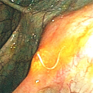
Figure A
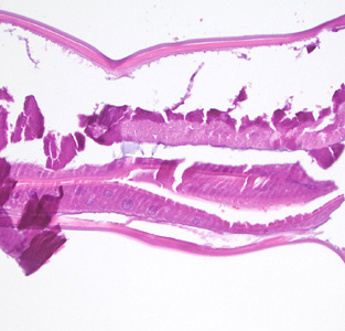
Figure D
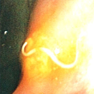
Figure B
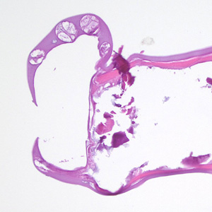
Figure E
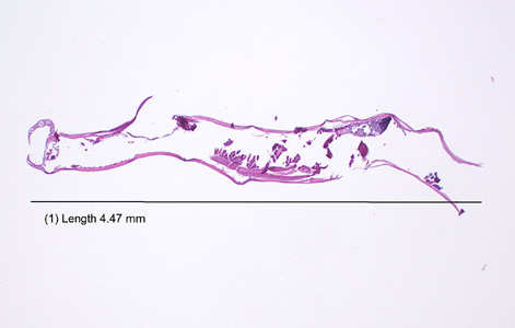
Figure C
This case and images were kindly provided by St. David’s South Austin Medical Center, Austin, TX.
