Case #436 – January, 2017
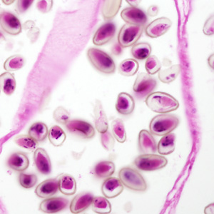
An immigrant from Korea presented with calculi in the intrahepatic biliary passages, cholangitis and early stage cholangiocarcinoma. Figures A–C show what was observed in a hematoxylin and eosin (H & E) stained liver tissue section. Figure A was captured at 40x magnification; Figures B and C at 200x magnification. The objects of interest in Figures B and C measured 30 micrometers on average. What is your diagnosis? Based on what criteria?

Figure A

Figure B

Figure C
Images presented in the dpdx case studies are from specimens submitted for diagnosis or archiving. On rare occasions, clinical histories given may be partly fictitious.