Case #457 – December, 2017
A nine-year-old female refugee from Eritrea with neutropenia and eosinophil counts within normal range had a stool examination as part of a refugee screening. Microscopic examination of an iodine wet mount preparation of a formalin-ethyl acetate (FEA) concentration of the stool sample submitted found what is shown in Figures A and B (taken at 400x) and C (at 1000x). What is your diagnosis? Based on what morphologic features?
This case and images were by provided by the Cadham Provincial Public Health Laboratory, Winnipeg, MB.

Figure A

Figure B

Figure C
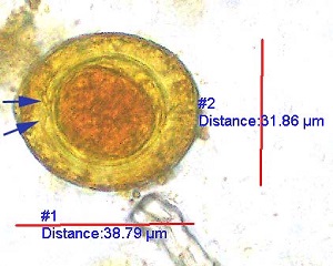
Figure A
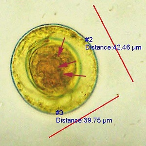
Figure B
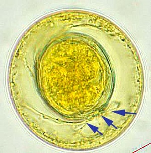
Figure C
This was a case of hymenolepiasis caused by Hymenolepis (Rodentolepis) nana (the dwarf tapeworm). Morphologic features shown included:
- Eggs within the size range for H. nana (30-50 micrometers).
- Presence of an oncosphere with hooklets, which are somewhat inconspicuous in image (red arrows; Figures B).
- Polar filaments between the inner and outer membranes (blue arrows, Figure A and C).
Infection has worldwide distribution and is most commonly seen in children
More on hymenolepiasis: https://www.cdc.gov/dpdx/hymenolepiasis/index.html
Images presented in the dpdx case studies are from specimens submitted for diagnosis or archiving. On rare occasions, clinical histories given may be partly fictitious.