Case #458 – December, 2017
A 69-year-old male, with recent travel to Peru, was undergoing a routine colonoscopy performed as a screen for colorectal cancer. The patient was asymptomatic and a stool ova and parasite examination (O & P) performed prior to the procedure was negative. However, the gastroenterologist recovered a worm from the ascending colon during the procedure and sent it to the pathology laboratory for identification. Figures A – C show what was observed on hematoxylin and eosin (H & E) stained slides. Eggs seen inside the worm measured 50 x 25 micrometers on average. What is your diagnosis? Based on what morphologic features.
This case and images were kindly provided by Keck Medical Center or University of Southern California.

Figure A

Figure B

Figure C

Figure A
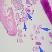
Figure B
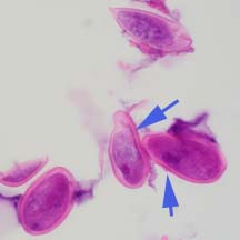
Figure C
This was a case of enterobiasis caused by Enterobius vermicularis (pinworm). Diagnostic morphologic features include:
- The cephalic expansions (black arrows, Figure A).
- Esophagus (purple arrows, Figure A) and anterior end of esophageal bulb (red arrow, Figure A).
- Eggs within the worm flattened on one side (blue arrows, Figures B and C) consistent with the species.
- Average egg size provided is within the range for E. vermicularius.
- Anatomical recovery site of the worm – the colon.
More on enterobiasis: https://www.cdc.gov/dpdx/enterobiasis/index.html
Images presented in the dpdx case studies are from specimens submitted for diagnosis or archiving. On rare occasions, clinical histories given may be partly fictitious.