Case #471 – July, 2018
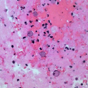
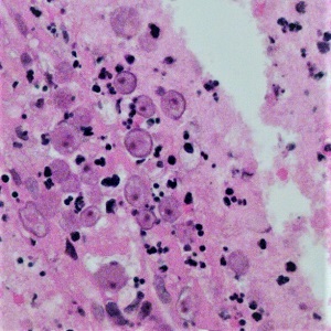
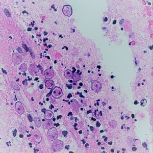
A 79 year-old man was diagnosed with encephalitis at a local hospital in Arizona. A Computed Tomography (CT) scan showed multiple brain lesions. A biopsy specimen from the occipital lobe was collected and sent to Pathology for routine histological workup. Images of structures shown in Figures A, B and C were observed by the pathologist on hematoxylin-and-eosin (H&E) stained sections and sent to DPDx for diagnostic assistance. The objects of interest shown in the Figures measured 10 – 25 µm in diameter. What is your diagnosis? Based on what criteria?
This case and the images were kindly contributed by the Health-Banner Desert Hospital, Mesa AZ

Figure A

Figure B

Figure C
Images presented in the dpdx case studies are from specimens submitted for diagnosis or archiving. On rare occasions, clinical histories given may be partly fictitious.

