Case #495 – July, 2019
A 68-year-old female with microscopic hematuria had a cystoscopy performed. A pathologist detected objects on hematoxylin and eosin (H & E) stained slides (Figures A–E) and on Papanicolaou stained slides of urine sediment (Figures F–I). A parasitic infection was suspected and images of the objects were captured and sent to DPDx for diagnostic assistance; the objects had a size range of 75-80 micrometers. What is your diagnosis? Based on what criteria?
This case was kindly provided by the Department of Pathology, Cork University Hospital, Ireland.
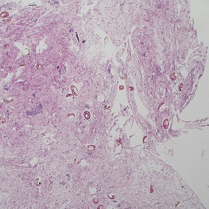
Figure A
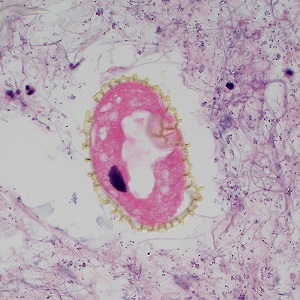
Figure B
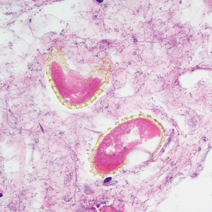
Figure C
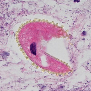
Figure D
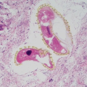
Figure E
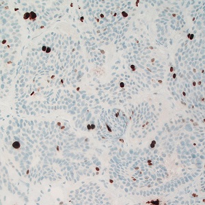
Figure F
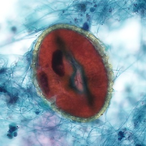
Figure G
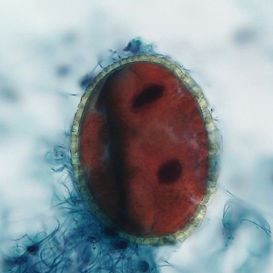
Figure H
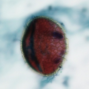
Figure I
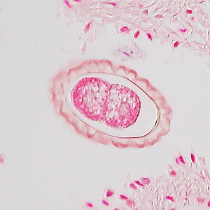
Figure J
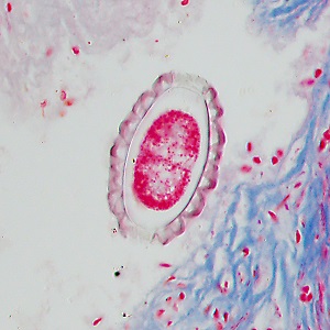
Figure K
Images presented in the dpdx case studies are from specimens submitted for diagnosis or archiving. On rare occasions, clinical histories given may be partly fictitious.